Saturday, July 29, 2006

Types of Dentition

1. Diphyodont. Most mammals--humans included--typically develope and erupt into their jaws two generations of teeth. The term literally means "two generations of teeth."
2. Monophyodont. Some mammals--such as the manatee, seals, and walruses have only a single generation of teeth.
3. Polyphyodont. Most reptiles and fishes develope a lifetime of generations of successional teeth--as if on a conveyer belt. Such teeth have a brief functional life and are anatomically simple in design.
4. Homodont. In many vertebrates, all of the teeth in the jaw are alike. They differ from each other only in size. The alligator is an example of homodontism.
5. Heterodont. Most mammals, humans included, develope distinctive classes of teeth that are regionally specialized.
6. Anodontia is the developmental absence of teeth. Among mammals, the whalebone whale and the anteater are toothless; their ancestors had teeth. In humans, anodontia is a pathological condition. Partial anodontia is one or a few teeth missing.

Dentition

Dentition is a term that describes all of the upper and lower teeth collectively.
Clinically, there are three dentitions.
The primary dentition consists of 20 teeth in all: ten upper and ten lower teeth. Primary teeth may also be called 'baby' teeth, deciduous, 'milk', or lacteal teeth. Primary teeth begin to appear at about age six months and are entirely replaced by about ages 12 - 13.
The mixed dentition is composed of both primary and permanent teeth. It commences with the eruption of the first of the permanent teeth at about age six, and ends with the loss of the last of the deciduous teeth at about the age of 12-13 years. The permanent dentition is composed of 32 teeth in all, 16 upper and 16 lower. Half of a dental arch (primary or permanent) is called a quadrant. The permanent teeth that replace deciduous teeth are call succedaneous teeth. (Succedaneous means literally, to replace. In dental science, permanent teeth that replace deciduous teeth are called successional teeth. Permanent molars, which replace nothing are called accessional teeth.)

Friday, July 28, 2006

dentation cusps

our teeth

dentition

i wanna continue with human dentition,lets take a look:

Tuesday, July 11, 2006

Forza ITALIA


Forza Azzurri
well, just like every other italian fan's, after 12 years, i'm very happy. congratulate to every one!!!!

man and monkey

Monday, July 03, 2006

cranium

" Elements of the Skull "
Skull: entire body framework of the head,including the lower jaw
Mandible: lower jaw
Cranium: skull without the mandible
Calvaria: cranium with out the face
Calotte: calvaria without the face
Splanchnocranium: facial skeleton
Neurocranium: braincase

" Bones of the Cranium "
Ethmoid
Floor of the cranium, inferior to the frontal bone and anterior to the sphenoid.Non-technically: Centre of the face, behind the nose.
Forms part of the nasal cavity and the orbits.Main support structure of the nasal cavity

Frontal
Forehead, extending down to form the upper surfaces of the orbits. Anterior roof of the skull.


Occipital
Back and base of the cranium, forms the back of the skull.Non-technically: Lower back of the head.
The occipital condyles (rounded surfaces at the base of the occipital bone) articulate with the atlas (first vertebra of the spine), enabling movement of the head relative to the spine.Has a large opening called the Foramen Magnus which the spinal cord passes through.

Parietal
Top and sides of the cranium, posterior roof of the skull.


Sphenoid
Anterior to the temporal bones and forms the base of cranium - behind the orbitals.Consists of a body, two "wings" and two "pterygoid processes" (not labelled on diagrams) that project downwards.
Articulates with the frontal, parietal and temporal bones.

Temporal
Sides of the skull, below the parietal bones, and above and behind the ears.
" Bones of the Face "
Hyoid
In the neck, below the tongue (held in place by ligaments and muscles between it and the styloid process of the temporal bone).
Supports the tongue, providing attachment sites for some tongue muscles, and also some muscles of the neck and pharynx.(Commonly fractured during strangulation, so studied in autopsies if strangulation suspected.)

Lacrimal
Behind and lateral to the nasal bone, also contribute to the orbits.(Smallest bones in the face.)
Contain foramina for the nasolacrimal ducts (tear ducts).

Mandible
Known as the lower jaw bone. Also forms the chin and sides of the face.(Largest, strongest facial bone.)
Bone into which the lower teeth are attached.The only moveable facial bone; motion of this bone is necessary for chewing food (the first stage of the digestion process).Each side of the mandible has a condyle and a coronoid process. The condyle articulates with the temporal bone to form the temporomandibular joint.

Maxilla
Upper jaw bone, which also forms the lower parts of the orbits.
Bone into which the upper teeth are attached.Each maxilla contains a maxillary sinus that drains fluid into the nasal cavity.

Nasal
Pair of small oblong bones that form the bridge and roof of the nose.


Palatine
Back of the roof of the mouth (hence not illustrated above). Small "L-shaped" bones.
Form the bottom of the orbitals and nasal cavities, and also the roof of the mouth.

Turbinator
Also known as Turbinate Bone and Nasal Concha. These terms refer to any of three thin bones that form the sides of the nasal cavity (not illustrated in the diagrams above).
Form the nasal cavities.

Vomer
Thin roughly triangular plate of bone on the floor of the nasal cavity and part of the nasal septum.
Separates the nasal cavities into left and right sides.

Zygomatic
Also known as Zygoma and Malar Bone.Commonly (non-medically) referred to as the Cheek Bone because it forms the prominent part of the cheeks. Also contributes to the orbits.

Directionel Terms


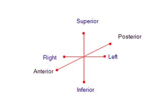
Directional Terms ,in osteology it is useful to refer the direction of motion or place ment of various skelton parts,most of these direction used for mammals not only human body. these experssion are used for the cranium.
  1. Superior: toward the head end of the homonid body.
  2. Inferior: opposide of the superior,for homonids,body parts away from the head,Caudal,toward a tail,is often used in the description of quadrupedal anatomy.
  3. Anterior: toward the front of homonid body.Ventral , toward the belly,may be used homologously for bipeds and quadrupedals.
  4. Posterior: opposite of the anterior,for homonids,toward the back of individual.Dorsal is often used for homologous parts of the quatruped anatomy.
  5. Medial: toward the midline.
  6. Lateral: opposit of the midline.away from the midline.
  7. Proximal: nearest the axial skeleton, usually used for limb bones.
  8. Distal: opposite of the proximal.farthest from the axial skeleton.
  9. External: outer.
  10. Internal: opposite of the external; inside.
  11. Endocranial: inner surface of the cranial valut .
  12. Ectocarnial: outer surface of the cranial valut.
  13. Superficail: close to surface
  14. Deep: opposite of the superficial, far from the surface.
  15. Subcutaneous: just below the skin.