Sunday, October 21, 2007

what are we going to do

friends sorry for long long breaks
as soon as posssible i will write again(nearly 1 month)
as you see we have just finished human skeleton and from now on i will start
new subject
Brain Endocasts
first i will share some information about brain system
our main reference is the human fossil record/volum 3/brain endocasts,the paleological evidence by ralph l.holloway
after brain we will disscus about DNA and then bio and evolution
and for the last sessionswe will talk about the use of this information in field anthropology.

Friday, April 06, 2007

just remeber

It is covinient to apply specific directional terms to foot just like hand.
Planter: refers to sole of the foot, its inferior surface in standard anatomical position.

Dorsal: opposite,superior surface.

Proximal/posterior: toward the tibia

Distal/anterior: twoard toe tips and the distalmost phalanges are reffered to as " terminal"

The Big Toe: somtimes called: Hallux
and its Ray is idetified as Ray1.
other rays are numbered as the hand.

Tarsal


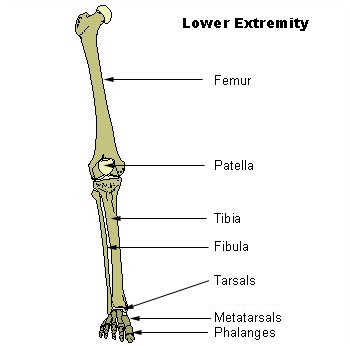
The 7 tarsal bone combine with the 5 metatarsals to form the longitudinal and traverse arches of the foot.

The talus articulates superiorly with the distal tibia and fibula at the ankle joint.

The Calcaneus forms the hell of the foot, supports talus and articulates anteriorly with the cuboid(3th largest tarsal bone)

The metatarsals articulate proximally with the cubid and three cuneiforms.

The 7th tarsal = the navicular, is interposed between the head of the talus and these cuneiforms.


Talus

called=Astragalus in other animals.

second largest in tarsal and is placed between the tibia and fibula superiorly and the calcaneus inferiorly.

no muscle attach to this bone.

It rests atop the calcaneus and articulates distally with the navicular.It forms the lower member of the talocrural joint.


tarsal parts are:

head

body

trochlea(saddle-shaped)

neck

groove for flexor hallucis longus

calcaneal (subtalar articular)

sulcus tali


Calcaneus

Heel bone

largest tarsal bone (second largest tarsal bone:talus/third:cuboid) + largest bone of the foot

it is located inferior to the talus and articulates anteriorly(distally) with the cuboid.


calcaneus parts are:

calcaneal tuber

lateral /medial processes

sustentaculum tali

sustentacular sulcus (groove)

peroneal tubercle


Cuboid

sits on the lateral side of the foot sandwiched between the calcaneus and fourth and fifth metatarsals,articulating with the navicular and 3th cuneiform.

it is recognized by its large size and projecting,pointed,proximal articular surface. it is the most cuboidal or cube-shaped,of the tarsal bones.

The cuboid tuberosity is a large tuberosity on the inferolateral surface of the bone.


Navicular

it is named for the strongly concave proximal surface that articulates with the head of the talus.

On the distal surface the navicular has a large facet divided by 2 ridges.

These set off the articular planes the 3 cuneiforms.

navicular often articulates with a large,blunt projection on the medial side of the bone.this tubercle is the main insertion of the Tibialis Posterior Muscle, a planter flexor of the foot and toes.


Medial (firsth)cuneiform

largest in 3 cuneiform.

sits between navicular and the base of the first metatarsal,articulating with these as well as the second cuneiform and the base of the MT2.It is the less wedge-shape than the other cuneiform and it is distinguished by the kidney-shaped facet for the base of the first metatarsal.


Intermediate (second) cuneiform

smallest of three cuneiforms.it is located between the navicula and second meta tarsal.also it articulates on either side with the first and third cuneiforms.


Lateral (third) cuneiform

it is the intermediate in size between the others,it is located in the center of the foot, articulating distally with the second,third and fourth metatarsals.medially it contacts the intermediate cuneform, laterally the cuboid, and proximally the navicular.


Tarsal ossifies from 1 center:(calcaneus is the exception) which has anepiphysis at its posterior end.

Most the tarsals are larger than carpals, they are more often recovered from archaeological sites.

foot


Foot's bones are obivousley the same as hand bones.
total bone's of foot(each): 26 (one less than each hand)
7 tarsal bones:
2 in proximal row
4 distal row
1 centered between rows
the tarsal are distally followed by a single row of 5 metatarsals
farther distally:
single row of 5 proximal phalanges,
single row of 4 intermediates phalanges,
single row of 5 distal/terminal phalanges.
(hand=)there are small Sesamoid bones that lie within tendons of the foot. in foot sesamoids is usually found below the head of first metatarsal.
elements of the foot skeleton :
tarsals
metatarsals
phalanges
The human foot has dramaticlly changed during its evolution from a grasping organ to a structure adapted to bipedal locomotion.
Most of the foot flexibity, and grasping abilities have been lost in humans as the foot had adapted to shock absorption and propulsion.

Friday, March 30, 2007

Knee

In humans the knee refers to the joints between the femur, tibia and patella. In quadrupeds, particularly horses and ungulates the term is commonly used to refer to the carpus, probably because of its similar hinge or ginglymus action. The joints between the femur, tibia and patella are known as the stifle in quadrupeds. In insects and other animals the term knee is used widely to refer to any ginglymus joint.
a comparison beteween bony anatomy of the human pelvic gridle and leg with that of our closest living realtives, the african apes,reveals profound evolutionary changes related to the acquisition of bipedality more than 4 million years.

Fibula


The long thin bone

Beside (lateral)tibia
Articulate twice with tibia,once with the talus
The main part in forming the lateral border of the ankle joint
Little wieght

Blood Supply
The blood supply is important for planning free tissue transfer because the fibula is commonly used to reconstruct the mandible. The shaft is supplied in its middle third by a large nutrient vessel from the peroneal artery. It is also perfused from its periosteum which receives many small branches from the peroneal artery. The proximal head and the epiphysis are supplied by a branch of the anterior tibial artery. In harvesting the bone the middle third is always taken and the ends preserved (4cm proximally and 6cm distally)

The fibula is ossified from three centers, one for the shaft, and one for either end.
Ossification begins in the body about the eighth week of fetal life, and extends toward the extremities. At birth the ends are cartilaginous.
Ossification commences in the lower end in the second year, and in the upper about the fourth year. The lower
epiphysis, the first to ossify, unites with the body about the twentieth year; the upper epiphysis joins about the twenty-fifth year.

the proximal and distal fibular ends are distinctive and are rarely confused with other bones.
fibular shafts are thin,straight and usually quatrilateral (sometimes rectangular) with sharp crests and corners.they are thus more irregular in cross section than either radial or ulnar shafts.

Saturday, March 24, 2007

TIBIA

The major weight-bearing bone of the lower leg
tibia articulates:

proximal: distal femur
twice lateral: fibula ( one proximal/one distal)
distal: talus( is called: astragalus in other animalsplace between fibula and tibia)
tibia parts are:
tibia plateau
medial condyle
lateral //
intercondylar eminence
medial intercondylar tubercle
lateral // //
superior fibular articulate facet
tibial tuberosity
tibial shaft
soleal (popliteal)lines
nutrient foramen
anterior surface (anterior crest)
medial surface
interosseus surface
// crest
medial malleolus
fibular notch
inferior fibular articulate surface
malleolar groove

Tibia ossifies from three center
shaft and two each end of the bone

Tibia shaft is mauch larger than ulnar or radial shafts the proximal and distal ends could be mistaken with proximal articular surface for the body of vertebra but the tibia articular surface is much smoother and denser.

Saturday, March 17, 2007

Patella


largest seamoid bone,articulated with the patellar surface of the distal femur(patellar notch). The patella rides in the Tendon the caudriceps femoris- the largest muscle of thigh and the primaay extensor of the knee. The patella functions to protect the knee joint, to lenghthen the lever arm of the quadricep, and to increase the area of contact between the patellar tendon and the femur.

Patella parts are:

Apex

Lateral articular facet

medial articular facet

the patella ossifies from a single center.

this bone might be mistaken for an os coxae fragment, but only in vary fragmentary state. The acetabulum of the os coxae is strongly hollowed, as opposed to the much flatter articular surface of the patella.

Patella is triangular in shape.its thin,pointed apex is distal, and the thicker, blunter end is proximal.

the lateral articular facet (articulates with the lateral condyle of the femur)= larger of the two facets.

Friday, March 16, 2007

Leg


Thursday, March 15, 2007

Leg


Leg: Femur,Patella,Tibia,Fibula
The evolution of the leg mirrors is like arm.The single thight bone, the femur, is the serial homolog of the upper arm bone,the humerus. Likewise, the lower bones of the leg, the tibia and fibula,are serial homolgs of the radius and ulna. the largest sesamoid bone in the body, the patella,lies at the knee joint. The bipedal locomotor mode practiced by homonids has resulted in major specializations of leg bones.
Femur
is the longest,heaviest,and strongest bone in the body.it supports all of the body's weight during standing,walking and running.
because of its strength and density, it is frequently recovered in forensic, archaeological and paleontological contexts. The femur is valuable bone because of the information it can provide on the stature of an individual.
the femur articulates with the acetabulum of the os coxae. Distally, it articulates with the patella and proximal tibia. The leg's actions at the hip include medial and lateral rotation, abduction, flexion and extension. At knee, motion is far more restricted,confined mostly to flexion and extension. Although the main knee action is that of a sliding hinge, this joint is one of the most complex in the body.
femur parts are:
head
spiral line
pectinel line
femoral shaft
linea aspera
nutrient foramen
medial supracondylar line (ridge)
popliteal surface
lateral condyle
lateral epicondyle
popliteal groove
medial condyle
intercondylar fossa (notch)
patellar surface

the femur ossifies from 5 centers: one for shaft, head,distal end,each trochanter.

the femoral head has a fovea (fossa) and is more complete sphere than the humeral head
the femoral shaft is larger,has a thicker cortex and is rounder in cross section than any other shaft. It has only one sharp corner,the linea aspera.

For isolated femoral heads the fovea is medial and displaced posteriorly and inferiorly. The posterinferior head-neck junction is more deeply excavated than anterosuperior junction.