Thursday, September 14, 2006

Scapula


It is a large, flat triangular bone with two basic surfaces, The posterior (dorsal) and the costal (anterior, or ventral). there are three borders that meet in three angles. The scapula articulates with the clavicle and the humerus.

The anterior (front) side of the scapula shows the fossa subscapularis (subscapular fossa) to which the subscapularis muscle attaches.
The posterior surface of the scapula is divided by a bony projection, the spina scapulae (opposite to the fossa subscapularis) into the supraspinous fossa and the infraspinous fossa. This projection is called the spine of the scapulae. It begins flat at the base of the shoulder bone, ascends in distal direction for all animals but carnivores and humans to its peak at about the middle of the scapula, this peak is called tuber scapulae. After this peak the spina scapulae steeply decays in height. For humans and carnivores and bovinae the spina runs into a forward pointing hook called acromion, which continues past the main part of the bone.
Another hook-like projection comes off the lateral angle of the scapula, and is called the coracoid process. The end of this hook is the site of attachment of many muscles, such as the coracobrachialis muscle.
Near the base of the coracoid process, so also on the lateral angle, there is a depression called the glenoid cavity. This forms the socket that the head of the humerus articulates with.


When fragmentary, the scapula might be mistaken for the pelvis. In all of its flat part, how ever the scapula is thinner than the pelvis. Indeed the scapular blade is mostly a single, thin layer of bone instead of spongy bone sandwiched between cortices as in the pelvis.

The fragment of scapular balde or infant scapulas could be mistaken for wings of the sphenoid, but the thin bone of the scapula is bounded by broken surfaces whereas broken sphenoid pieces normally have free or sutural edges.


Wednesday, September 13, 2006

Appendicula Skeleton: Shoulder Gridle 1: Clavicle


The depature of fishes from their aquatic habitat brought profound changes in locomotion. The forelimb gridle ( shoulder gridle), attached to the rear of head, detached and moved tailward, leaving a flexible neck. As this gridle moved back, some of the head's dermal armor and gill muscles remained attached to it, and our own shoulder gridle is still attached to the skull by derivatives of some of these primitive gill muscles. The remaining dermal bone element constitutes part of our clavicle. The human shoulder gridle provides support and articulation for tht humerus and anchors a variety of muscles. The clavicle's function az a strut for the shoulder is made obvious when fracture of this bone is accompanied by anteromedial collaps of the shoulder. The shoulder gridle embraces the Thorax posteriorly, laterallym and anteriorly, providing a platform for movements of the forelimb.
Clavicle
The clavicle forms the anterior portion of the shoulder girdle. It is a long bone, curved somewhat like the italic letter f, and placed nearly horizontally at the upper and anterior part of the thorax, immediately above the first rib.Medially, it articulates with the manubrium of the sternum(breast-bone) at the sternoclavicular joint. At its lateral end it articulates with the acromion of the scapula (shoulder blade) at the acromioclavicular joint.
  • It is shorter, thinner, less curved, and smoother in women than in men.
It has a rounded medial end and a flattened lateral end.
The clavicle serves several functions:
It serves as a rigid support from which the scapula and free limb are suspended. This arrangement keeps the upper limb away from the thorax so that the arm has maximum range of movement.
Covers the cervicoaxillary canal (passageway between the neck and arm), through which several important structures pass.
Transmits impacts from the upper limb to the axial skeleton.
Even though it is classified as a long bone, the clavicle has no medullary (bone marrow) cavity like other long bones. It is made up of spongy (cancellous) bone with a shell of compact bone. The clavicle bone is the only bone that does not have marrow. It is a dermal bone derived from elements originally attached to the skull.

Monday, September 11, 2006

Male or Female? Pelvis


Differences between the Male and Female Pelves:


(male pelvis.left)
  • I think pelvis is the main part in human skeleton that we use for showing male or female.
( female pelvis .up. right)


The female pelvis is distinguished from that of the male by its bones being more delicate and its depth less. The whole pelvis is less massive, and its muscular impressions are slightly marked. The ilia are less sloped, and the anterior iliac spines more widely separated; hence the greater lateral prominence of the hips. The preauricular sulcus is more commonly present and better marked. The superior aperture of the lesser pelvis is larger in the female than in the male; it is more nearly circular, and its obliquity is greater. The cavity is shallower and wider; the sacrum is shorter wider, and its upper part is less curved; the obturator foramina are triangular in shape and smaller in size than in the male. The inferior aperture is larger and the coccyx more movable. The sciatic notches are wider and shallower, and the spines of the ischia project less inward. The acetabula are smaller and look more distinctly forward . The ischial tuberosities and the acetabula are wider apart, and the former are more everted. The pubic symphysis is less deep, and the pubic arch is wider and more rounded than in the male, where it is an angle rather than an arch.
The size of the pelvis varies not only in the two sexes, but also in different members of the same sex, and does not appear to be influenced in any way by the height of the individual. Women of short stature, as a rule, have broad pelves. Occasionally the pelvis is equally contracted in all its dimensions, so much so that all its diameters measure 12.5 mm. less than the average, and this even in well-formed women of average height. The principal divergences, however, are found at the superior aperture, and affect the relation of the antero-posterior to the transverse diameter. Thus the superior aperture may be elliptical either in a transverse or an antero-posterior direction, the transverse diameter in the former, and the antero-posterior in the latter, greatly exceeding the other diameters; in other instances it is almost circular.


Pelvis

The pelvis (pl. pelvises or pelves) is the bony structure located at the base of the spine (properly known as the caudal end). The pelvis incorporates the socket portion of the hip joint for each leg (in bipeds) or hind leg (in quadrupeds). It forms the lower limb (or hind-limb) girdle of the skeleton.

The pelvis is symmetrical and each side is actually made up of three separate bones

  • the upper half (the broad "wings") is the ilium
  • the middle (the top half of the lower "loops") is the pubis
  • the bottom (the lower half of the "loops") is the ischium
    These three bones fuse together with age and are collectively known as the hip bone, os coxa, or the innominate bone. The pelvis is joined to the sacrum bone by ligaments (the sacroiliac joint), and the hip bones nest in specially shaped sockets (the acetabulum) on each side. The upper edge of the ilium is known as the iliac crest. The place at the front of the pelvis where the two sides join together is called the symphysis pubis. This is normally a very inflexible joint, but it softens and becomes more flexible during late pregnancy, allowing it to expand during labour for the baby's head to pass through.
Position of the Pelvis:In the erect posture, the pelvis is placed obliquely with regard to the trunk: the plane of the superior aperture forms an angle of from 50° to 60°, and that of the inferior aperture one of about 15° with the horizontal plane. The pelvic surface of the symphysis pubis looks upward and backward, the concavity of the sacrum and coccyx downward and forward. The position of the pelvis in the erect posture may be indicated by holding it so that the anterior superior iliac spines and the front of the top of the symphysis pubis are in the same vertical plane.

Coccyx


The vestigial guman tail,is highly variable in shape, with three to five (most often four) variably fused segments. The rudimentary vertebrae of coccyx show articular and transverse process superiorley,but t hey lack pedicles, laminae, and spinous processes. The sacral articulation is via the superior coccygeal body as well as a relatively large pair of tubercles called the Cornau. The latter are rudimentary articular processes that contact the sacrum. The coccyx may fuse with the sacrum late in life.
As with the sacrumm the individual vartebral elements of the coccyx decrease in size inferiorly, and horizonyal lines of fusion can be seen between adjacent coccylgeal vertebrae. The coccyx pelvic muscles and ligaments.

female or male? Sacrum

Differences in the Sacrum of the Male and Female
In the female the sacrum is shorter and wider than in the male; the lower half forms a greater angle with the upper; the upper half is nearly straight, the lower half presenting the greatest amount of curvature. The bone is also directed more obliquely backward; this increases the size of the pelvic cavity and renders the sacrovertebral angle more prominent. In the male the curvature is more evenly distributed over the whole length of the bone, and is altogether greater than in the female.

Saturday, September 09, 2006

some pic


dorsal surface

base of Sacrum


The sacral and coccygeal vertebræ:
consist at an early period of life of nine separate segments which are united in the adult, so as to form two bones, five entering into the formation of the sacrum, four into that of the coccyx. The sacral vertebrae fuse during adolesence into one immoble, wedgeshaped bone,the sacrum. This bone is typically formed from five segments but sometimes is made of four or six.
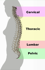
location: at the base of the vertebral column and articulates on the left and right with the two Os Coxae and inferiorly with the small Coccyx.

The sacrum: (fig.1)
is a large, triangular bone at the base of the spine and at the upper and back part of the pelvic cavity, The sacrum is a large,triangular bone at the base of the spine and at the upper and back part of the pelvic cavity, where it is inserted like a wedge between the two hip bones. Its upper part or base articulates with the last lumbar vertebra, its apex with the tailbone or coccyx.
It is curved upon itself and placed obliquely. It is concave facing forwards, thus its curvature is considered a
kyphosis. The base projects forward as the sacral promontory internally, and articulates with the last lumbar vertebra to form the prominent sacrovertebral angle. The central part is directed backward, so as to give increased capacity to the pelvic cavity.
The name is derived from the
Latin sacer, "sacred", a translation of the Greek hieron (osteon), meaning sacred or strong bone. This is supposedly derived from the belief that it could not be destroyed and was the part that would allow rising from the dead.

pelvic surface


Pelvic Surface (facies pelvina):

The pelvic surface ( fig. up) is concave from above downward, and slightly so from side to side. Its middle part is crossed by four transverse ridges, the positions of which correspond with the original planes of separation between the five segments of the bone. The portions of bone intervening between the ridges are the bodies of the sacral vertebræ. The body of the first segment is of large size, and in form resembles that of a lumbar vertebra; the succeeding ones diminish from above downward, are flattened from before backward, and curved so as to accommodate themselves to the form of the sacrum, being concave in front, convex behind. At the ends of the ridges are seen the anterior sacral foramina, four in number on either side, somewhat rounded in form, diminishing in size from above downward, and directed lateralward and forward; they give exit to the anterior divisions of the sacral nerves and entrance to the lateral sacral arteries. Lateral to these foramina are the lateral parts of the sacrum, each consisting of five separate segments at an early period of life; in the adult, these are blended with the bodies and with each other. Each lateral part is traversed by four broad, shallow grooves, which lodge the anterior divisions of the sacral nerves, and are separated by prominent ridges of bone which give origin to the Piriformis muscle.
If a sagittal section be made through the center of the sacrum the bodies are seen to be united at their circumferences by bone, wide intervals being left centrally, which, in the fresh state, are filled by the intervertebral fibrocartilages. In some bones this union is more complete between the lower than the upper segments.

Lateral Surface:

The lateral surface is broad above(fig. 1), but narrowed into a thin edge below. The upper half presents in front an ear-shaped surface, the auricular surface, covered with cartilage in the fresh state, for articulation with the ilium. Behind it is a rough surface, the sacral tuberosity, on which are three deep and uneven impressions, for the attachment of the posterior sacroiliac ligament. The lower half is thin, and ends in a projection called the inferior lateral angle; medial to this angle is a notch, which is converted into a foramen by the transverse process of the first piece of the coccyx, and transmits the anterior division of the fifth sacral nerve. The thin lower half of the lateral surface gives attachment to the sacrotuberous and sacrospinous ligaments, to some fibers of the Glutæus maximus behind, and to the Coccygeus in front.


Dorsal Surface (facies dorsalis):

The dorsal surface is convex and narrower than the pelvic. In the middle line it displays a crest, the middle sacral crest, surmounted by three or four tubercles, the rudimentary spinous processes of the upper three or four sacral vertebræ. On either side of the middle sacral crest is a shallow groove, the sacral groove, which gives origin to the Multifidus, the floor of the groove being formed by the united laminæ of the corresponding vertebræ. The laminæ of the fifth sacral vertebra, and sometimes those of the fourth, fail to meet behind, and thus a hiatus or deficiency occurs in the posterior wall of the sacral canal. On the lateral aspect of the sacral groove is a linear series of tubercles produced by the fusion of the articular processes which together form the indistinct sacral articular crests. The articular processes of the first sacral vertebra are large and oval in shape; their facets are concave from side to side, look backward and medialward, and articulate with the facets on the inferior processes of the fifth lumbar vertebra. The tubercles which represent the inferior articular processes of the fifth sacral vertebra are prolonged downward as rounded processes, which are named the sacral cornua, and are connected to the cornua of the coccyx. Lateral to the articular processes are the four posterior sacral foramina; they are smaller in size and less regular in form than the anterior, and transmit the posterior divisions of the sacral nerves. On the lateral side of the posterior sacral foramina is a series of tubercles, which represent the transverse processes of the sacral vertebræ, and form the lateral crests of the sacrum. The transverse tubercles of the first sacral vertebra are large and very distinct; they, together with the transverse tubercles of the second vertebra, give attachment to the horizontal parts of the posterior sacroiliac ligaments; those of the third vertebra give attachment to the oblique fasciculi of the posterior sacroiliac ligaments; and those of the fourth and fifth to the sacrotuberous ligaments.

Base (basis oss. sacri):

The base of the sacrum, which is broad and expanded, is directed upward and forward. In the middle is a large oval articular surface, the upper surface of the body of the first sacral vertebra, which is connected with the under surface of the body of the last lumbar vertebra by an intervertebral fibrocartilage. Behind this is the large triangular orifice of the sacral canal, which is completed by the laminæ and spinous process of the first sacral vertebra. The superior articular processes project from it on either side; they are oval, concave, directed backward and medialward, like the superior articular processes of a lumbar vertebra. They are attached to the body of the first sacral vertebra and to the alæ by short thick pedicles; on the upper surface of each pedicle is a vertebral notch, which forms the lower part of the foramen between the last lumbar and first sacral vertebræ. On either side of the body is a large triangular surface, which supports the Psoas major and the lumbosacral trunk, and in the articulated pelvis is continuous with the iliac fossa. This is called the ala; it is slightly concave from side to side, convex from before backward, and gives attachment to a few of the fibers of the Iliacus. The posterior fourth of the ala represents the transverse process, and its anterior three-fourths the costal process of the first sacral segment.

Pelvic Gridle: Sacrum, Coccyx and Os Coxae

The bony structure at the base of the front limbs is the shoulder gridle and the one at the base of the hind limbs is the Pelvic gridle. The pelvic gridles of terrestial vartebrates are connected to the vertebral column and are much larger than their homologs in fish. These adaptation are required for weight bearing and muscle attachment in terrestial forms. In early land-dwelling vertebrates, the right and left limb gridles joined dorsally with the sacral vertebrae to form a bony ring around the rear of the trunk.
The adult human bony pelvic comprises three main elemnts: the right and left Os Coxae and the Sacrum and Coccyx. The sacrum and coccyx are part of the axial skeleton and are actually variably fused vartebrae. The bony pelvis functions to support and protect the abdominal and pelvic organs. In additon, it anchor muscles of the abdomen leg. Unlike the shoulder gridle, which is movable platform, the pelvic gridle is firmly fixed to the axial skeleton via its vertebral element, the sacrum.

The pelvis is divided by an oblique plane passing through the prominence of the sacrum, the arcuate and pectineal lines, and the upper margin of the symphysis pubis, into the greater and the lesser pelvis. The circumference of this plane is termed the linea terminalis or pelvic brim.

The Greater or False Pelvis (pelvis major):

The greater pelvis is the expanded portion of the cavity situated above and in front of the pelvic brim. It is bounded on either side by the ilium; in front it is incomplete, presenting a wide interval between the anterior borders of the ilia, which is filled up in the fresh state by the parietes of the abdomen; behind is a deep notch on either side between the ilium and the base of the sacrum. It supports the intestines, and transmits part of their weight to the anterior wall of the abdomen.

The Lesser or True Pelvis (pelvis minor):

The lesser pelvis is that part of the pelvic cavity which is situated below and behind the pelvic brim. Its bony walls are more complete than those of the greater pelvis. For convenience of description, it is divided into an inlet bounded by the superior circumference, and outlet bounded by the inferior circumference, and a cavity.

Friday, September 08, 2006

lateral view of the Vertebral Column

the Thorax from infront

different regions of the Vertbral column

Thursday, September 07, 2006

Thorax and sternum

In primitive, air-breathin are fishes, breathing was accomplished by sawllowing movements in which air was gulped into the lungs. Early reptiles improved on this system when they evolved a means to respire via the muscuskeletal machanics of the thoracic skeleton. this was made possible as the thoracic ribs extended ventrally from the vertebrae column to reach the sternum. The sternum formed akind of ventral bony column that fused into a bony bar and anchored the distal ends of the ribs. Further soft tissue specializations led to more sophisticated breathing function.
Thorax
Or chest, is like basket or cage composed of cartilage and bone. It is attached dorsally to the vertbral column. It is also the base ehich upper limbs are attached. The major bones forming the thorax are the sternum and twelve ribs on each side.
Male or Female
The thorax of the female differs from that of the male as follows:
1. Its capacity is less.
2. The sternum is shorter.
3. The upper margin of the sternum is on a level with the lower part of the body of the third thoracic vertebra, whereas in the male it is on a level with the lower part of the body of the second.
4. The upper ribs are more movable, and so allow a greater enlargement of the upper part of the thorax.
Sternum
Or breastbone, function at its upper end to connect the shoulder gridle ( Clavicle and Scapula ) to the thorax.The sternum is an elongated, flattened bone, forming the middle portion of the anterior wall of the thorax. its margins articulate with the cartilages of the first seven pairs of ribs. It consists of three parts, named from above downward, the manubrium, the body or gladiolus, and the xiphoid process. The bone is composed of 3 main parts in adulthood but develops from six segements. This segment joints may all fuse in adulthood, but their location is indicated by the costal notches along each side of the sternum. Its average length in the adult is about 17 cm., and is rather greater in the male than in the female.

Ribs

There are usually twelve ribs on each side of the thorax, for a total of twenty-four in adults. The number of ribs is variable,but this number may be increased by the development of a cervical or lumbar rib so there may be eleven or thirteen on a side. The upper ribs (1 - 7) atriculate directly with the sides of the sternum via cartilage.They are connected behind with the vertebral column. Ribs 8,9 and 10 are interconnected medially by common cartilages that attach to the sternum. The last two ribs, 11 and 12, have free floating ventral ends. all ribs articaulte via their proximal ends with thoracic vertebrae. The ribs usually increase in length from rib 1 to rib 7 and decrease from rib 7 to rib 12.
For studing ribs there are several sidings like:
  1. Head : the swollwn proximal part of the rib
  2. Neck : The short segment between the head and the rib's articulation with the transverse of the thoracic vertebrae
  3. Tubercle : located on the posteroinferior corner of each ribs.
  4. Angle : sharve curve in the bone lsteral to tubercle.
  5. Shaft : the curved, tapering segment between the tubercle and the rib's distal (ventral)end.
  6. Costal Groove : the grrove along the medial side of the inferior edge of the rib shaft.
  7. Sternal End : the anterior (ventral) end of the shaft. Its surface change substantially with increasing the age.
  8. Cranial (upper) Edge: for most of the ribs is blunt, smooth and convex.
  9. Caudal (lower) Edge : for most of the ribs is sharp, with a costal groove on the medial surface.

The first seven ribs with the sternum are called true or vertebro-sternal ribs, the remaining five are false ribs.

Special ribs are : 1,2,10,11 and 12th.

for further information and photoes take a look at http://www.bartleby.com/107/28.html

Names of the Individual Vertebrae

Individual vertebrae named according to region and position, from superior to inferior
Cervical – 7 vertebrae (C1-C7)
Thoracic – 12 vertebrae (T1-T12)
Lumbar – 5 vertebrae (L1-L5)
Sacral – 5 (fused) vertebrae (S1-S5)
Coccygeal – 3-5 vertebrae (Co1-Co5)

Skeleton's Parts

The skeleton has two parts:
the Axial skeleton and the Appendicular skeleton.
The axial skeleton includes:

the skull, the hyoid bone, the vertebral column (spine, sacrum, and coccyx), the sternum, and the ribs. Its components are aligned along the long axis of the body.
The appendicular skeleton includes:

the bones of the upper extremities (arms, forearms, and hands), the pectoral (shoulder) girdle, the pelvic (hip) girdle, and the bones of the lower extremities (thigh, knee, leg, and foot). Its components are outside the body main axis.

Axial Skeleton

The vertebral column has three groups of vertebrae and two sets of fused bones. These vertebrae include seven cervical (neck) vertebrae, twelve thoracic (upper back) vertebrae, and five lumbar (lower back) vertebrae. Five fused vertebrae form the sacrum and from three to five fused small vertebrae form the coccyx (tail bone). The vertebrae form a column of bone that protects the spinal cord. The thoracic vertebrae have facets (indentations) upon their surfaces that articulate (meet) with the ribs.
Curves
The cervical curve, convex forward, begins at the apex of the odontoid (tooth-like) process, and ends at the middle of the second thoracic vertebra; it is the least marked of all the curves. The thoracic curve, concave forward, begins at the middle of the second and ends at the middle of the twelfth thoracic vertebra. Its most prominent point behind corresponds to the spinous process of the seventh thoracic vertebra. The lumbar curve is more marked in the female than in the male; it begins at the middle of the last thoracic vertebra, and ends at the sacrovertebral angle. It is convex anteriorly, the convexity of the lower three vertebrae being much greater than that of the upper two. The pelvic curve begins at the sacrovertebral articulation, and ends at the point of the coccyx; its concavity is directed downward and forward.
The thoracic and pelvic curves are termed primary curves, because they alone are present during fetal life.In the early eymbro, the vertebral column is C-shaped, and the cervical and lumbar curvatures are not yet present in a newborn infant.
The cervical and lumbar curves are compensatory or secondary, and are developed after birth, the former when the child is able to hold up its head (at three or four months), and to sit upright (at nine months), the latter at twelve or eighteen months, when the child begins to walk.
True or False
The vertebrosternal (true) ribs are the first seven ribs; they are "true" because they attach directly to the sternum (breast bone). Ribs eight through twelve are the false ribs because they indirectly attach to the sternum or they lack a sternal attachment. Ribs eight through ten are the vertebrochondral ribs because they attach indirectly to the sternum by cartilage. Ribs eleven and twelve are called floating (vertebral) ribs because they do not attach to the sternum.
Hyoid Bone
In the middle line below the chin can be felt the body of the hyoid bone,- hyoid bone is very importanat for following the human language evolution- just below which is the prominence of the thyroid cartilage called "Adam's apple," better marked in men than in women.